 Swine Flu
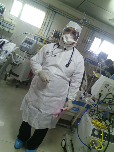
 took a heavy toll if lives in Rajasthan specially Jaipur last winter. Statistically highest number of death were reported from this city. We bring you a first hand account of the conditions as prevailed in SMS hospital Jaipur written by its senior dcotor incharge of Swine Flu there:
Just the very the thought of taking charge of all the three dedicated swine flu ICU’s of our hospital spelt excitement. I was heading a team of 4 Assistant Professors, 4 Senior Residents, and 18 Junior Residents. I thought to myself. I will be able to help people in misery. Little did I know what was in store for me? From seeing young men and women coming with sore throat and breathlessness to getting incubated. We felt helpless as their PaO2 and SpO2 decreased. I was overwhelmed. I would like to share my experience from the day of hospital admission from symptoms, history, physical examination, labs to treatment it was a herculean task. I would like to thank all my colleagues who worked day and night to contain and combat this deadly disease.
We admitted Category C patients in our ICU. They had complaints of breathlessness, chest pain, drowsiness, fall in BP, sputum mixed with blood, cyanosis, children with influenza-like illness with red flag signs. “Red flag signs included somnolence, high and persistent fever, inability to feed well, convulsions, shortness of breath, difficulty in breathing and worsening of underlying condition.
On admission, in our hospital specimens were taken from nasopharynx and nasal cavity for confirmation of diagnosis of H1N1.
Symptoms at presentation included cough, fever, breathlessness, bluish discoloration of lips, and nail beds..31% (6) of the patients were on ventilator at the time of admission. 32% (6) of the patients were cyanosed and 37% were breathless at the time of presentation. The average day of presentation after onset of symptoms in patients requiring ventilator was 6th day with 74% of the patients presenting between day 4 and day 10 when antiretroviral therapy was also initiated. Antiretroviral therapy was not initiated within 48 h of onset of symptoms in any patient. A detailed medical history and examination done at the time of admission, their time of presentation, and co morbidities were recorded. Majority of patients requiring ventilation on admission were referred from other centers.
H1N1 infection had multisystem involvement of respiratory, cardiac, neurological, gastrointestinal, and renal systems. Presence of risk factors increased the severity of disease and altered the prognosis. Risk factors included infants and young children, pregnant women, asthma, COPD, congestive cardiac failure, diabetes, chronic renal disease, chronic hepatic disease, Neuromuscular, Neurocognitive, Seizure Disorders, Hemoglobinopathies, Immune-Suppression, HIV Infection, Immunosuppressive Medication, Malignancy, children receiving chronic aspirin therapy, persons aged 65 years and older, Hypertension, Hypothyroidism.
Physical ExaminationA detailed examination done at the time of admission showed that in most patients there was evidence of hypotension. On inspection there was evidence of breathing difficulty. On auscultation there was evidence of Crepts.
All the laboratory investigations in the form of chest X-ray, hemogram, arterial blood gas (ABG) analysis, serum electrolytes, blood sugar, renal and liver function tests, and endotracheal aspirate and blood culture results, which were done at the time of admission or subsequently, were noted. Diagnosis of H1N1was confirmed in all the patients.
Repeat cultures were performed at weekly intervals. X-ray chest showed bilateral pulmonary infiltrates in all the patients. Review of all the investigations done revealed that 8 out of 19 patients, developed extra-pulmonary organ dysfunction during the course of disease.15% of the patients developed deranged renal status with raised levels of blood urea and serum creatinine. In patients with endotracheal tubes in situ, endotracheal aspirate was taken. Electrocardiography, echocardiography, and Creatine kinase levels were checked to determine cardiac involvement in patients with worsening dyspnea or prolonged weakness. 26% of our ventilated patients had cardiac involvement as evident on ECG in the form of ST-elevation with Q wave. 5 out of 19 patients eventually required high inotropic support to maintain hemodynamic.
Treatment of swine-flu positive patients requiring ventilatory supportAll patients requiring ventilatory support had significant respiratory involvement in the form of pneumonia, acute Respiratory Distress Syndrome (ARDS) and respiratory failure. Patients were treated on the basis of severity of disease, presentation characteristics, diagnostic findings, treatment modalities, and the final outcome. Mean age of patients requiring ventilator support was 30 years with minimum age of 2 years.
All patients were given pharmacological treatment with oral oseltamivir. Pharmacological treatment was initiated at the time of admission with all adult patients receiving oseltamivir 75 mg BD through nasogastric tube. One 12 kg, 2-year-old child was treated with oral oseltamivir 30 mg BD...Patients with severe pneumonia and acute respiratory failure were given ventilatory support. ABG analysis was performed twice daily in patients on mechanical ventilation, as per the protocol.
The patients given ventilator support were either cyanosed or breathless, with average SpO2 82% at the time of initiation of ventilatory therapy. Invasive mechanical ventilation is preferred mode of ventilation over noninvasive one as noninvasive ventilation can worsen the outcome. Unlike most of the patients with ARDS, these patients are young with severe refractory hypoxemia which is difficult to manage. All patients were paralyzed and ventilated as per ARDS protocol with ventilator set to maintain plateau pressure less than 30 cm H2O.
Besides low tidal volume mechanical ventilation, various strategies were employed to improve oxygenation in these patients. These patients were given PEEP along with prolonged inspiratory phase and higher FiO2. Our patients were managed with permissible PEEP, low tidal volume, and high respiratory rate keeping in mind the oxygenation and plateau pressure goals. We tried a few novel approaches which we had heard of, eg prone positioning improves oxygenation by optimizing lung recruitment and ventilation – perfusion matching. Positive pressure mechanical ventilation was the mainstay of our treatment though it had some disadvantages.
We used the ventilation strategies. Fluid conservative therapy decreases lung edema. Cell based therapy with allogeneic human MSCs has emerged as a promising approach to therapy. Supportive management included intravenous fluids, vasopressors for shock and paracetamol/ibuprofen for fever or myalgia. Empirical antibiotic therapy was initiated in all the ventilated patients after obtaining endotracheal aspirate and blood cultures. Third generation cephalosporin’s were administered in all patients with dose alteration as per the renal function. Cultures were positive for bacterial infections in 4 out of 19 ventilated patients, and the antibiotic therapy was remodeled as per the sensitivity reports. 15% of our ventilated patients also went into acute renal shutdown. Patients also had CNS manifestations in the form of unconsciousness, altered mental status, seizures, and confusion.14 out of 19 patients expired in swine-flu ICU. The majority of patients who expired were of the age group of 20–39 years.
Despite the best efforts of our government to prevent the occurrence of disease, and early diagnosis/intervention a large number of these cases developed a severe form of the disease in the early days of outbreak. 35% of hospitalized patients required ventilatory support based on the condition at the time of presentation, ABG reports and SpO2levels. As pregnancy was one of the major co morbid factor and poor nutritional and immunological status of females in our country, there were more female patients requiring ventilatory support with female: male ratio being 1.7:1.The interval from onset of symptoms to initiation of antiretroviral therapy correlates maximally with the severity of the disease. Benefit of antiretroviral therapy has strongest evidence when it is started within 48 h after onset of symptoms. The average day of presentation of patients to our health care facility was between 4th and 10thdays and antiretroviral therapy was initiated then. All patients had respiratory involvement in the form of breathlessness, cyanosis, and hypoxia as indicated by pulse Oximetry or ABG analysis.. All patients who required ventilatory support as per the protocol were managed in the lines of ARDS guidelines.
To conclude, my experience of patients with swine-flu is that they should be isolated and managed aggressively. The prognosis of the disease is best when treatment is started as early as 48 hrs after onset of symptoms. Co morbidities increase the risk of death in ventilated patients. The earliest signs of deterioration of the respiratory parameters warrant early intervention with ventilatory support, antiviral therapy, and good supportive treatment. A Critical Care wing should be started in all Tertiary Care Hospitals dedicated solely for Infectious Diseases. As Stress levels amongst Doctors working in this Unit is very high their duties should be arranged accordingly. Close coordination between all specialties be it Anesthesia, Medicine, Chest and TB or Pathology is very important. Retrospective data of all the ventilated patients revealed that antiretroviral therapy was not initiated within 48 hrs of onset of symptoms in any of the patient due to delayed reporting to a hospital with the required facility. This may have contributed to increasing the severity of illness.
Dr.Tarun Lall,
Professor, Department of Anesthesia,
SMS Hospital, Jaipur
 Swine Flu took a heavy toll if lives in Rajasthan specially Jaipur last winter. Statistically highest number of death were reported from this city. We bring you a first hand account of the conditions as prevailed in SMS hospital Jaipur written by its senior dcotor incharge of Swine Flu there: